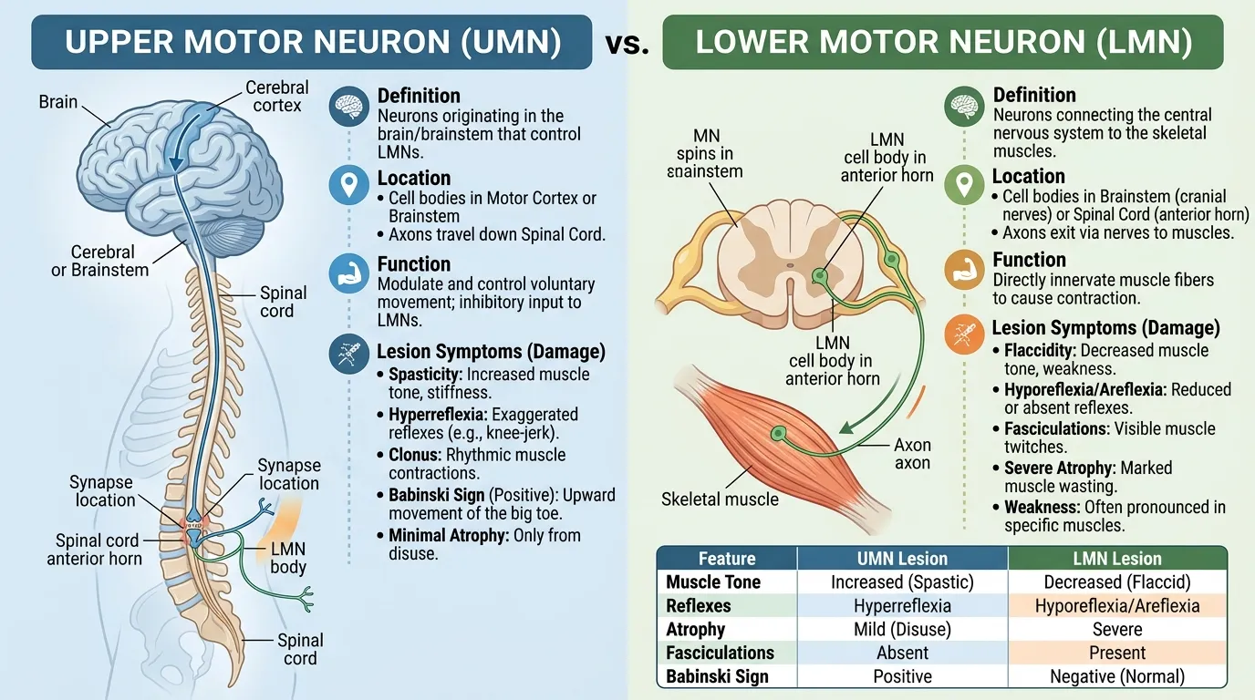
UMN vs LMN refers to the clinical differences between upper motor neuron and lower motor neuron lesions. UMN lesions typically cause weakness, increased muscle tone, hyperreflexia, and an extensor Babinski response. LMN lesions typically cause weakness with decreased tone, reduced or absent reflexes, muscle atrophy, and fasciculations.
Key Takeaways
- UMN means upper motor neuron; LMN means lower motor neuron.
- UMN lesions commonly produce spasticity and hyperreflexia.
- LMN lesions commonly produce flaccidity, hyporeflexia, atrophy, and fasciculations.
- A positive Babinski sign strongly suggests corticospinal tract dysfunction.
- Acute severe spinal cord injury can temporarily produce flaccidity before typical UMN signs develop.
What Are UMNs and LMNs?
The easiest way to understand UMN vs LMN is to think about the motor pathway as a chain.
The upper motor neuron carries movement-related signals from the brain toward the brainstem or spinal cord. It then influences the lower motor neuron. The lower motor neuron provides the final connection to skeletal muscle and directly helps produce muscle contraction.
Upper Motor Neurons
Upper motor neurons are located within the central nervous system. They include neurons involved in pathways that descend from the motor areas of the brain to the spinal cord or brainstem.
The corticospinal tract is particularly important for voluntary movement. Damage along this pathway can produce the characteristic UMN pattern of weakness, increased tone, and abnormal reflexes.
Lower Motor Neurons
Lower motor neurons are the final motor pathway connecting the nervous system to skeletal muscle. Their cell bodies can be found in the anterior horn of the spinal cord or in motor cranial nerve nuclei, with their axons traveling toward muscles.
Damage to a lower motor neuron or its associated peripheral pathway can interrupt the signal reaching the muscle. This commonly results in reduced tone, reduced reflexes, muscle wasting, and sometimes fasciculations.
UMN vs LMN: Key Differences
The biggest differences are seen during a neurological examination.
| Feature | UMN Lesion | LMN Lesion |
| Muscle tone | Increased | Decreased |
| Typical tone pattern | Spasticity | Flaccidity |
| Deep tendon reflexes | Increased | Decreased or absent |
| Babinski response | Often extensor/positive | Usually absent or normal |
| Muscle atrophy | Usually less prominent initially | Often prominent |
| Fasciculations | Generally absent | Common |
| Weakness | Often follows a pyramidal pattern | Often follows a nerve, root, or segmental pattern |
| Clonus | May occur | Generally absent |
These findings are useful because they help clinicians localize where along the motor pathway a problem may be occurring.
UMN Lesion Signs
An upper motor neuron lesion affects descending motor pathways and changes the normal control of spinal reflex circuits.
Muscle Tone and Spasticity
One of the classic UMN findings is increased muscle tone, often described as spasticity.
A person may feel unusually stiff, and movement of an affected limb can encounter increased resistance. Merck Manual identifies increased tone and spasticity as important clues to UMN dysfunction.
For example, someone with a corticospinal tract lesion may have difficulty moving a leg smoothly because the muscles are unusually stiff.
Reflexes and Babinski Sign
UMN lesions commonly cause hyperreflexia, meaning deep tendon reflexes are more brisk than expected. Clonus can also occur.
Another important finding is the Babinski response. In adults, an abnormal extensor plantar response—where the big toe moves upward when the sole is stimulated—can indicate corticospinal tract dysfunction.
Weakness and Muscle Bulk
UMN lesions cause weakness, but the pattern can be different from peripheral nerve damage. Weakness may follow a pyramidal distribution, depending on the location of the lesion.
Muscle wasting can eventually occur because of reduced use, but marked early neurogenic atrophy is more characteristic of LMN damage.
LMN Lesion Signs
A lower motor neuron lesion directly affects the motor pathway that connects with skeletal muscle.
Flaccidity and Reduced Reflexes
LMN dysfunction commonly produces decreased muscle tone, giving the affected limb a soft or floppy quality.
Deep tendon reflexes are generally reduced or absent because the lower motor neuron is part of the reflex pathway itself.
For example, a peripheral nerve injury can interfere with both voluntary movement and the reflex involving muscles supplied by that nerve.
Muscle Atrophy and Fasciculations
One of the clearest LMN clues is muscle atrophy.
When the nerve supply to a muscle is significantly impaired, the muscle can lose bulk over time. LMN lesions may also produce fasciculations, which are small, visible involuntary muscle twitches.
A combination of weakness, reduced reflexes, low tone, atrophy, and fasciculations strongly suggests an LMN pattern, although the exact diagnosis requires clinical evaluation.
Common Causes and Clinical Examples
UMN and LMN patterns can occur in many different neurological disorders.
Examples of conditions associated with UMN findings include stroke, multiple sclerosis, and spinal cord lesions affecting corticospinal pathways.
LMN findings can occur with disorders involving the anterior horn cells, nerve roots, peripheral nerves, or other parts of the motor unit. Peripheral neuropathies and certain motor neuron disorders can produce LMN features.
Importantly, the clinical pattern alone does not establish a specific disease. Doctors combine the neurological examination with the patient’s history and, when appropriate, tests such as MRI, nerve conduction studies, and electromyography.
Important Exception: Acute UMN Lesions
A common source of confusion when learning UMN vs LMN is that an acute UMN lesion does not always immediately look like a classic UMN lesion.
For example, severe acute spinal cord injury can initially cause spinal shock, characterized by flaccid weakness and reduced reflexes. Over the following days or weeks, the examination may evolve toward spasticity and hyperreflexia.
This is why timing matters when interpreting neurological signs. A patient examined shortly after an acute injury may not yet show the textbook UMN pattern.
How Clinicians Distinguish UMN From LMN Lesions
Clinicians generally look at several findings together rather than relying on one sign.
They may assess:
- Muscle strength — identifies weakness and its distribution.
- Muscle tone — increased tone suggests an UMN pattern, while decreased tone suggests an LMN pattern.
- Deep tendon reflexes — brisk reflexes favor UMN dysfunction; reduced or absent reflexes favor LMN dysfunction.
- Plantar response — an extensor Babinski response supports corticospinal tract dysfunction.
- Muscle bulk — marked neurogenic atrophy points toward LMN involvement.
- Fasciculations — visible muscle twitching can support LMN involvement.
- Distribution of weakness — the pattern can help localize the affected part of the nervous system.
The combination of findings is more informative than any single feature.
FAQs:
Is Babinski positive in UMN or LMN lesions?
A positive or extensor Babinski response is associated with upper motor neuron dysfunction, particularly corticospinal tract involvement. It is not the typical response expected from an LMN lesion.
Is spasticity a UMN or LMN sign?
Spasticity is classically an UMN sign. It reflects increased muscle tone associated with disruption of descending motor control. LMN lesions instead usually produce reduced tone or flaccidity.
Is muscle atrophy UMN or LMN?
Marked, neurogenic muscle atrophy is more characteristic of an LMN lesion. UMN lesions can eventually cause some muscle wasting from disuse, so atrophy by itself does not always identify the lesion location.
Can a condition cause both UMN and LMN signs?
Yes. Some neurological disorders can affect both motor neuron levels. Amyotrophic lateral sclerosis (ALS) is a classic example because it can produce both UMN findings, such as spasticity, and LMN findings, such as atrophy and fasciculations.
Conclusion
Understanding UMN vs LMN becomes much easier when you focus on the pattern of neurological findings. UMN lesions typically produce increased tone, hyperreflexia, spasticity, and an extensor plantar response, while LMN lesions more often produce decreased tone, reduced reflexes, muscle atrophy, and fasciculations.
The important exception is acute severe spinal cord injury, where early findings can temporarily resemble an LMN pattern before typical UMN signs develop.
For students, the fastest way to remember the distinction is: UMN = spasticity + hyperreflexia; LMN = flaccidity + hyporeflexia, with atrophy and fasciculations often present.