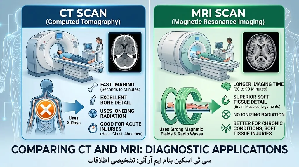
CT and MRI are different medical imaging tests, and neither is always better. CT uses X-rays and is usually faster, making it useful for emergencies, trauma, bones, bleeding, and many lung or abdominal problems. MRI uses a strong magnetic field and radio waves, does not use ionizing radiation, and generally provides better soft-tissue detail.
Key Takeaways
- CT scans use X-rays, while MRI uses magnets and radio waves.
- CT is generally faster and is commonly used when doctors need answers quickly.
- MRI does not use ionizing radiation and is especially useful for detailed soft-tissue imaging.
- CT can be particularly useful for fractures, trauma, bleeding, lungs, and complex anatomy.
- MRI is often preferred for ligaments, tendons, cartilage, nerves, the spinal cord, and many brain conditions.
CT vs MRI: What Is the Difference?
CT stands for computed tomography. It uses X-rays and computer processing to create detailed cross-sectional images, or “slices,” of the body. CT can show bones, blood vessels, organs, and other tissues and can produce detailed images very quickly.
MRI stands for magnetic resonance imaging. Instead of X-rays, MRI uses a powerful magnetic field and radio waves to create images. MRI is especially valuable when doctors need strong contrast between different types of soft tissue.
The important point is that CT and MRI are complementary tests. A doctor chooses between them based on what needs to be seen, how urgently an answer is needed, and the patient’s medical situation.
How a CT Scan Works
During a CT scan, X-ray equipment rotates around the body while a computer processes the information into detailed images. A CT examination can be completed very quickly, which is one reason it is widely used in emergency care.
How an MRI Works
MRI uses a strong magnetic field and radiofrequency energy to produce detailed images of internal structures. It does not use ionizing radiation. Because MRI examinations can require the patient to remain still for longer periods, they are generally slower than CT examinations.
CT vs MRI: Side-by-Side Comparison
| Feature | CT Scan | MRI |
| Full name | Computed tomography | Magnetic resonance imaging |
| Main technology | X-rays | Magnetic field + radio waves |
| Ionizing radiation | Yes | No |
| Speed | Usually faster | Usually slower |
| Bone imaging | Excellent | Useful, but CT is often preferred for detailed bone anatomy |
| Soft-tissue detail | Good | Generally excellent |
| Emergency use | Very common | Less commonly the first choice when speed is critical |
| Metal-device concerns | Fewer MRI-specific restrictions | Important because of the strong magnetic field |
| Common uses | Trauma, fractures, bleeding, lungs, abdominal problems | Brain, spine, joints, nerves, muscles, soft tissues |
The exact choice depends on the body part and clinical question. There is no single scan that is best for every situation.
When Is a CT Scan Usually Used?
CT is particularly useful when physicians need fast, detailed images.
Emergency and Trauma
After a serious accident or injury, doctors may need to quickly determine whether there is internal bleeding, a fracture, or organ damage. CT can rapidly provide this information and is widely used in emergency settings.
For example, after a major car accident, a CT may help doctors evaluate several areas of the body without requiring a lengthy examination.
Bones, Lungs, and Internal Injuries
CT is excellent for showing complex bone anatomy and can also provide detailed information about the lungs, blood vessels, and many internal organs.
It may be used to investigate problems such as:
- Complex fractures
- Chest and lung abnormalities
- Internal injuries
- Abdominal problems
- Blood vessel conditions
- Certain types of tumors
Brain Emergencies
A brain CT is often used when speed matters, such as after a head injury or when a stroke is suspected. It can quickly identify problems such as bleeding, swelling, and certain fractures.
When Is an MRI Usually Used?
MRI becomes particularly useful when doctors need high-detail images of soft tissues.
Soft-Tissue Problems
MRI can distinguish different types of soft tissue very well. This makes it useful for evaluating muscles, tendons, ligaments, cartilage, nerves, and many organs.
For example, an MRI may be ordered when a doctor suspects an ACL tear, rotator cuff injury, cartilage problem, or certain spinal conditions.
Brain and Spine Conditions
MRI can provide highly detailed images of the brain and spinal structures. It is commonly used when doctors need more detailed information about brain tissue than a CT can provide.
For some types of stroke, MRI can be particularly sensitive, while CT may be preferred initially when emergency speed is critical.
Muscles, Joints, and Ligaments
MRI is often useful for sports injuries and other musculoskeletal problems. It can help visualize structures such as:
- Ligaments
- Tendons
- Cartilage
- Muscles
- Nerves
- Spinal structures
Johns Hopkins notes that MRI is particularly useful for conditions such as meniscus tears, ACL injuries, Achilles tendon injuries, and rotator cuff tears.
CT vs MRI for the Brain
Both CT and MRI can be used to examine the brain, but they serve different purposes.
CT is often favored when doctors need a rapid assessment, such as after a head injury or when brain bleeding is suspected.
MRI generally provides greater detail of brain tissue and may be used for conditions involving tumors, certain strokes, dementia-related changes, and other neurological problems.
So, if someone asks, “Is MRI always better for the brain?” the answer is no. The appropriate test depends on the medical question and how quickly an answer is needed.
CT vs MRI: Radiation and Safety
One of the biggest differences is radiation.
CT uses ionizing radiation because it uses X-rays. The amount depends on the type of CT examination and the body area being scanned. Medical imaging providers use radiation doses appropriate for obtaining the needed diagnostic information.
MRI does not use ionizing radiation. Instead, it uses a powerful magnetic field and radio waves.
However, MRI is not automatically risk-free. The magnetic environment creates important safety considerations for certain metal objects, implants, and electronic medical devices. Patients should tell the MRI team about implants, devices, or metal in the body before the examination.
Pregnancy can also affect the imaging decision. Doctors consider whether CT radiation is necessary and whether another examination, such as MRI or ultrasound, can provide the information needed.
CT vs MRI: Contrast Material
Sometimes a scan is performed with contrast material to make certain structures easier to see.
CT commonly uses iodine-based contrast when contrast is needed. MRI may use a contrast agent called gadolinium for certain examinations. Contrast is not required for every CT or MRI.
If you have kidney disease, a previous reaction to contrast, or another relevant medical condition, tell your healthcare team before the examination.
Which Scan Takes Longer?
CT is generally much faster than MRI. This difference can be important when a patient is seriously injured or when doctors need information immediately. CT examinations can acquire images rapidly, while MRI examinations often require more time and the patient must remain still.
The exact duration varies by body part, scanner, protocol, and whether contrast is required.
Is CT or MRI Better?
There is no universal winner between CT and MRI.
A CT may be more appropriate when speed, bone detail, trauma assessment, or rapid detection of bleeding is important. MRI may be more appropriate when doctors need detailed soft-tissue information or want to avoid ionizing radiation.
The right examination is the one that best answers the specific medical question.
FAQs:
Is MRI safer than CT?
MRI does not use ionizing radiation, which is a major advantage over CT. However, MRI has its own safety considerations because of its powerful magnetic field. Certain implants and devices require special evaluation before an MRI.
Which is better for detecting tumors?
It depends on the tumor and body area. MRI often provides excellent soft-tissue detail and can be very useful for evaluating tumors, while CT can also be important for detecting and staging many cancers. The appropriate test depends on the suspected condition and the clinical question.
Can you have an MRI instead of a CT?
Sometimes, but not always. CT and MRI provide different types of information. In an emergency, CT may be chosen because it is faster. In other situations, MRI may provide information that CT cannot show as clearly.
Does an MRI use radiation?
No. MRI does not use ionizing radiation. It creates images using a strong magnetic field and radio waves.
Why would a doctor choose CT first?
CT is often selected when doctors need rapid imaging, particularly in emergency situations. It can quickly show internal bleeding, fractures, organ injuries, and other urgent problems.
Conclusion
CT vs MRI is not really a question of which scan is always better. CT is fast and highly useful for emergencies, trauma, bones, bleeding, lungs, and complex anatomy. MRI takes longer but provides excellent soft-tissue detail and does not use ionizing radiation.
Your healthcare professional chooses the imaging test based on your symptoms, the body part being examined, how quickly the information is needed, and your individual safety considerations.